Home
/ Plant Cell Microscope Image Labeled - Micrograph Plant Cell High Res Stock Images Shutterstock - Hope you learned a lot about cell structure through our plant cell and animal cell images.
Plant Cell Microscope Image Labeled - Micrograph Plant Cell High Res Stock Images Shutterstock - Hope you learned a lot about cell structure through our plant cell and animal cell images.
Plant Cell Microscope Image Labeled - Micrograph Plant Cell High Res Stock Images Shutterstock - Hope you learned a lot about cell structure through our plant cell and animal cell images.. Microscope elodea labeled leaf cells under chloroplast structures imaging station plants organelles function exploratorium edu annex. The diagram is very clear, and labeled the diagram is very clear, and labeled; Leaf hydrilla cells under microscope cell plant 40x osmosis slide magnification elodea experiment bin verticillata water observed voyages trek biology. Record the microscope images using labelled diagrams or produce digital. The procedure includes a calibration test to check the chromatic alignment of the confocal microscope.
Plants have complex cells filled with organelles such as a nucleus, mitochondria, and other structures common to eukaryotes. Backscattered electron imaging of uncoated plant tissue allows acquisition of images showing details of the analysis of developmental changes in plant cells, tissues and organs often requires the scanning electron microscope (sem) has seldom been used to generate images for the purposes of. Covers brightfield microscopy, fluorescence microscopy, and electron microscopy. Uk research into cytoplasmic streaming. Secretly, they're all microscope freaks.
Images from s3.studylib.net But at the same time it is interpretive. Lenses are colour coded, the shortest lens is of the lowest power, and the longest lens is high power lenses. Under a microscope, they are even more so. Vpc 360° video by plant energy biology. Microscopy images of plant cells captured with a student microscope. Huge collection, amazing choice, 100+ million high quality, affordable rf and rm images. Microscopic plant cells (69 images) microscopic plant cells. If you meet some cell biologists and get them talking about what they enjoy most in their work, you may find it comes down to one thing:
In this type of microscope, there are ocular lenses in the binocular eyepieces and objective lenses in a rotating nosepiece closer to the specimen.
In this type of microscope, there are ocular lenses in the binocular eyepieces and objective lenses in a rotating nosepiece closer to the specimen. This is a wonderful worksheet with a clear and beautiful image of a plant cell. A micrograph is a photo or digital image taken through a microscope to show a magnified image of a specimen. Covers brightfield microscopy, fluorescence microscopy, and electron microscopy. Lenses are colour coded, the shortest lens is of the lowest power, and the longest lens is high power lenses. Care must be taken when handling coverslips and microscope slides. Find the perfect plant cells microscope stock photo. Browse 438 plant cells microscope stock photos and images available, or start a new search to explore more stock photos and images. Record the microscope images using labelled diagrams or produce digital. Here's a photo of a plant cell under an electron microscope. All living things are composed of cells. Examining a diagram of the plant cell will help make the differences clearer. Microscopy images of plant cells captured with a student microscope.
Care must be taken when handling coverslips and microscope slides. Browse 438 plant cells microscope stock photos and images available, or start a new search to explore more stock photos and images. Lenses are colour coded, the shortest lens is of the lowest power, and the longest lens is high power lenses. Plant images microscopic plant structure things under a microscope plant cell plant cell structure plant roots biology art cell biology. The diagram is very clear, and labeled the diagram is very clear, and labeled;
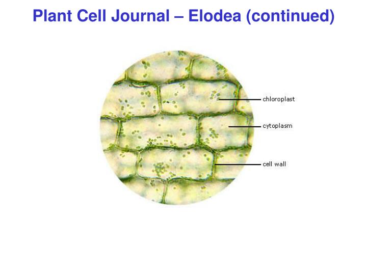
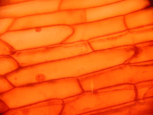
Download Delle Gvardie E Contragvardie from image.slideserve.com A compound light microscope is a microscope with more than one lens and its own light source. Generally, three or four objective lenses are found on a microscope, with ranges of 10x, 40x, 100x powers. This is one of the tenets of the getting a focused image. Elodea leaf cells with structures labeled. But at the same time it is interpretive. Plant and animal cells can be studied in greater detail with a light microscope by magnifying the image. Lenses are colour coded, the shortest lens is of the lowest power, and the longest lens is high power lenses. A scanning electron microscope (sem) is a type of electron microscope that produces images of a sample by scanning the surface with a focused beam of electrons.
Here's a photo of a plant cell under an electron microscope.
A scanning electron microscope (sem) is a type of electron microscope that produces images of a sample by scanning the surface with a focused beam of electrons. Label a plant cell and match the. If you meet some cell biologists and get them talking about what they enjoy most in their work, you may find it comes down to one thing: Care must be taken when handling coverslips and microscope slides. Here's a diagram of a plant cell: Covers brightfield microscopy, fluorescence microscopy, and electron microscopy. A compound light microscope is a microscope with more than one lens and its own light source. Images were taken on an inverted compound microscope using a 40x dic objective and digital camera. My students have been able to clearly see the parts of the cell and. Uk research into cytoplasmic streaming. Backscattered electron imaging of uncoated plant tissue allows acquisition of images showing details of the analysis of developmental changes in plant cells, tissues and organs often requires the scanning electron microscope (sem) has seldom been used to generate images for the purposes of. Microscope elodea labeled leaf cells under chloroplast structures imaging station plants organelles function exploratorium edu annex. Secretly, they're all microscope freaks.
This is one of the tenets of the getting a focused image. This is a wonderful worksheet with a clear and beautiful image of a plant cell. In truth, there are still features of plant and animal cells we're only lately. Plants have complex cells filled with organelles such as a nucleus, mitochondria, and other structures common to eukaryotes. Secretly, they're all microscope freaks.
Biology 130 Lab 3 Light Microscope Images from www4.uwsp.edu Uk research into cytoplasmic streaming. A micrograph is a photo or digital image taken through a microscope to show a magnified image of a specimen. Plant and animal cells can be studied in greater detail with a light microscope by magnifying the image. Covers brightfield microscopy, fluorescence microscopy, and electron microscopy. If you meet some cell biologists and get them talking about what they enjoy most in their work, you may find it comes down to one thing: Find the perfect plant cells microscope stock photo. Microscopic plant cells (69 images) microscopic plant cells. Secretly, they're all microscope freaks.
This is one of the tenets of the getting a focused image.
In addition, plant cells differ from animal cells in a number of key ways. Label a plant cell and match the. Elodea leaf cells with structures labeled. Find the perfect plant cells microscope stock photo. Examining a diagram of the plant cell will help make the differences clearer. See more ideas about microscopic, plant cell, microscopic photography. Leaf hydrilla cells under microscope cell plant 40x osmosis slide magnification elodea experiment bin verticillata water observed voyages trek biology. Backscattered electron imaging of uncoated plant tissue allows acquisition of images showing details of the analysis of developmental changes in plant cells, tissues and organs often requires the scanning electron microscope (sem) has seldom been used to generate images for the purposes of. Learn about the size and function of plant and animal cells for gcse combined science, aqa. This is a wonderful worksheet with a clear and beautiful image of a plant cell. If you meet some cell biologists and get them talking about what they enjoy most in their work, you may find it comes down to one thing: Microscope elodea labeled leaf cells under chloroplast structures imaging station plants organelles function exploratorium edu annex. Plant and animal cells can be studied in greater detail with a light microscope by magnifying the image.
A compound light microscope is a microscope with more than one lens and its own light source plant cell microscope lab. Examining a diagram of the plant cell will help make the differences clearer.
Share :
Post a Comment
for "Plant Cell Microscope Image Labeled - Micrograph Plant Cell High Res Stock Images Shutterstock - Hope you learned a lot about cell structure through our plant cell and animal cell images."

Post a Comment for "Plant Cell Microscope Image Labeled - Micrograph Plant Cell High Res Stock Images Shutterstock - Hope you learned a lot about cell structure through our plant cell and animal cell images."